Blog

Normal Anatomy of the Knee Joint A Comprehensive Guide by Dr. Vivek Bansal
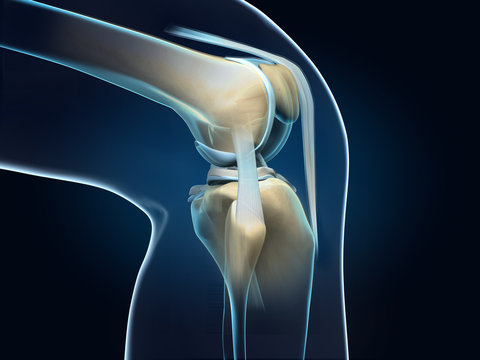
The knee joint is one of the most complex and vital joints in the human body. It supports almost all activities involving leg movement, including walking, running, and jumping. Understanding the normal anatomy of the knee joint is essential for diagnosing and treating various conditions and injuries.
Key Components of the Knee Joint
- Bones The knee joint is a hinge joint formed by the interaction of three main bones:
- Femur (thigh bone): The largest bone in the body, it extends from the hip to the knee.
- Tibia (shin bone): It runs from the knee to the ankle and forms the lower part of the knee joint.
- Patella (kneecap): This small, triangular bone sits in front of the knee joint and helps protect the joint.
- Cartilage Cartilage is a smooth, rubbery tissue that covers the ends of the bones in the knee. It helps reduce friction during movement and absorbs the shock placed on the knee.
- Articular Cartilage: This type of cartilage covers the ends of the femur, tibia, and back of the patella, allowing smooth movement within the joint.
- Menisci: These are two crescent-shaped pieces of cartilage (medial and lateral meniscus) that sit between the femur and tibia. The menisci act as shock absorbers and provide stability to the joint.
- Ligaments Ligaments are strong bands of tissue that connect bones to each other, providing stability to the knee joint. There are four major ligaments in the knee:
- Anterior Cruciate Ligament (ACL): Prevents the tibia from sliding out in front of the femur and provides rotational stability.
- Posterior Cruciate Ligament (PCL): Works with the ACL to control the back-and-forth motion of the knee.
- Medial Collateral Ligament (MCL): Runs along the inside of the knee and prevents excessive sideways movement.
- Lateral Collateral Ligament (LCL): Positioned on the outer side of the knee, it resists outward forces.
- Tendons Tendons connect muscles to bones, facilitating movement. The two key tendons in the knee are:
- Quadriceps Tendon: Connects the quadriceps muscle in the front of the thigh to the patella.
- Patellar Tendon: Connects the patella to the tibia, aiding in leg extension.
- Muscles Several muscles contribute to the knee’s movement:
- Quadriceps: A group of four muscles located at the front of the thigh, responsible for straightening the knee.
- Hamstrings: Located at the back of the thigh, these muscles help bend the knee.
- Bursae The knee joint contains small fluid-filled sacs called bursae. These sacs reduce friction and allow smooth movement between tissues, particularly in areas with significant movement.
Function of the Knee Joint
The knee joint plays a crucial role in supporting body weight and enabling lower-limb mobility. Its unique structure allows for:
- Flexion and Extension: The primary motion of the knee is bending (flexion) and straightening (extension), which is essential for walking, running, and climbing.
- Rotation: While primarily a hinge joint, the knee also allows for a small degree of rotation, particularly when the knee is bent.
Attri
0